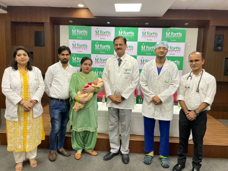
Noida, 22nd September 2022: Doctors at Fortis Hospital Noida successfully treated a one-day-old newborn, with a rare condition of brain fluid-filled swollen lump on the neck (cervical meningomyelocele). The lump had a high risk of rupture, if not operated on immediately. A team of doctors led by Dr Rahul Gupta, Director, Neurosurgery, Fortis Noida operated on the newborn, and in a span of 3 hours, removed the swollen lesion from the neck.
The baby was born at a hospital in Ghaziabad and later shifted to Fortis Noida on the same day, as the Ghaziabad Hospital was not equipped to handle such complex cases. The baby presented with an unusually large swelling on the neck, which was tense and looked like an inflated balloon. The doctors first dressed the swelling to prevent any skin injury. A cervical spine and brain MRI was conducted which showed part of the baby’s cervical spinal cord going into the swollen area. The condition was diagnosed as cervical meningomyelocele and in view of the large size of the swelling and risk of rupture, the doctors decided to perform the surgery at the earliest to remove the fluid-filled sac. In most conditions, such lumps are formed owing to congenital malformation which occurs in the first 4 weeks of life inside the womb. Some studies suggest deficiency of folic acid in mother’s body as the cause.
Giving details of the surgery, Dr Rahul Gupta, Director, Neurosurgery, Fortis Hospital, Noida said, “This is a very rare condition and it is also very difficult to perform the surgery owing to the risk factors while operating on a one-day-old newborn. There was a high risk in giving anaesthesia to the baby and the dosage had to be very precisely administered. We took the help of Dr Varun Jain, HOD, Neuro Anaesthesia, Fortis Hospital Noida, who inserted an intravenous cannula and conducted the MRI under sedation. The baby’s bones were very thin and soft, so after opening the swelling, we could see the spinal cord adherent to the inner cavity of the sac. We separated the adhesions with great difficulty, under maximum magnification of the operating microscope. Dural closure after repositioning the spinal cord was another challenge. All the tissues (bones, muscles and ligaments were very tiny and a high-end operating microscope was needed to magnify them. Neural tissue (spinal cord) was separated from surrounding adherent tissues and put back into the spinal canal (normal space for the spinal cord in the bony spine). Since there is a risk of leakage of brain fluid from the wound, meticulous closure of different layers of the wound was done. Any brain fluid leak in the post-operative period could have derailed the whole effort as the cervical region has more chances of complications.”


